Featured post

Take a Quick Quiz

Loading...

Saturday, 17 March 2018

      The activity of this blog is now totally shifted to the new growing website : eMedExam.com

Monday, 4 December 2017

Seven Tips to Make a Professional Patient Interview


Whether you are a medical student or a practicing physician, it's always a challenge when you face a patient coming with few symptoms (in non-emergency setting), and seeking for your help.
It's almost fruitless to skip history taking towards lab investigations or imaging.

The following are history taking tips that if you follow, you are likely to explore your patient's symptoms very efficiently, and makes your life easier:

1- Make sure you, and your patient are in comfort (both physical and psychological). You both should have enough time to talk, and listen to each other, in a calm office.

2- You should make a short introduction, in such a way that you win the patient's trust. All barriers should be removed, so the patient feels free to tell you everything. This can be by asking about his/her job, shortly discussing the media news, finance, cultural events, daily life, ..etc. You should be so friendly.

3- At the beginning let the patient talk freely, without much interruption. Ask open questions and stay listening. An example of  open questions, is "Can you tell me about your problem?". Let him/her say everything he/she likes to tell. Only interrupt when the patient's words deviate from the main focus of the interview, to bring back the direction of the conversation in the medical history taking general path.


4- When it seems that the patient has nothing more to add, start changing your questions to closed questions. An example of closed questions, is "how many times you vomited yesterday?".
The aim of closed questions is to close the gaps in the history. 
Remember that when the patient tells the story, it won't be perfect. There must be many holes that to be sealed. When you can identify these holes, you can seal them with very specific and targeted closed questions.

5- When a patients tells you something that cannot be easily believable, you should confirm by further asking, probably again and again. You have always confirm what the patient mean, especially if the answer is a critical piece of information.

6- During history taking, make the habit of ongoing symptom analysis. I mean by analysis here, is to translate symptoms into "pathology or sometimes a diagnosis". For example, productive cough, breathlessness, and pleuritic chest pain can be "translated" into lobar pneumonia.
This requires training and adequate basic knowledge of the differential diagnosis of each symptom alone and in combination with other symptoms.

7- By the end of your history taking you should have now analyzed the clinical data enough to make a list of reasonable differential diagnoses. This list should start with the most likely, and ends with the least likely diagnosis. In this way, you are now comfortable enough to proceed towards the diagnostic steps, and you are almost certainly near to make a definitive diagnosis.


Thursday, 5 October 2017

How to use mind maps in your study of medicine?


Medical students are totally aware of the difficulty in recalling all what they study.

Perhaps, each one has his/her preferred method of exam preparation, but in general, the study methods don’t significantly differ.


Some of us prefer mnemonics, and some depend on their “extraordinary” memorizing talent.

But, is it possible for everyone to build an efficient storage memory, with little of effort and more fun?


The answer is yes. You can use the technique of mind maps.

Mind mapping is a very well know method of drawing thoughts on paper to make them clear, and easy to remember. Mind maps can be simple explanatory diagrams or a large detailed charts.

They should be eye catching, and readily understood, and it’s preferable to contain simple drawings, colours, words or phrases, and logic arrangement of all these elements.


So, how can we use mind maps in our medical study?


To make things clear, here is an example:


Of course, the above shown diagram is just an example. It simply shows the possible complications of peptic ulcer disease.


Mind maps may be made in any way, with as little or as much images, or words, depending on personal preferences.

Everyone has his/her own style of mind mapping. Even the details in the map can vary.


You may ask: it’s OK now, I can remember the details about acute pancreatitis, but there are hundreds of subjects in medicine, and if I create a map for each, I will get totally lost.

So how can I solve this?


First, it’s still easy. Mind maps were invented to help memorization. You don’t need to build a map for a subject that you know very well.


Second, don’t repeat the same map pattern for each subject as much as you can. Make each map peculiar. You may put even pictures or write words that are not related to medicine but can enable you to memorize without difficulty.


Your aim is to draw a map that’ s not forgettable and easily recallable.

Put your effort to make some link between the subject title and the map elements, if possible.


If you start with this method right now, you will notice, by time, that your ability to efficiently memorize your subject, is steadily improving, and longer lasting , with a lot of fun.


Friday, 14 April 2017

Sarcoidosis short notes


Sarcoidosis is a granulomatous multi-system disease of unknown etiology.
It usually affects younger no middle aged, with a the disease being more severe in African blacks.

The typical histopathological picture is non-caseating  granulomas, with heroes the presence of epitheliod cells, T lymphocytes and macrophages.

Commonly affected organs are the lungs, skin, and eyes.
Symptoms of lung disease are non-productive cough and dyspnea. Cor pulmonale may develop.
There may be pulmonary infiltrates, an less commonly pulmonary fibrosis.
Chest examination is usually normal. Fine crepitations may be present. Finger clubbing is rare , and if present it should raise the suspicion of an alternative diagnosis.

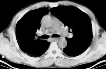
The triad of arthralgias, bihilar lymphadenopathy, and erythema nodosum is known as Löfgren’s sindrome.

Diagnosis is suggested by symptoms and signs. Biopsy from enlarged lymph nodes, skin, or transbronchial biopsy are indicated.


Pulmonary function tests help assess the disease severity and to monitor response to treatment. Abnormalities may be a restrictive lung defect with decrease in gas transfer.

Serum angiotensin-converting enzyme (ACE) is usually raised, but it has low specificity, and is not of diagnostic value.
Treatment is not indicated with normal lung parenchyma (even if there is hilar lymphadenopathy), and pulmonary function tests.

Patients with affected lung parenchyma or abnormal pulmonary function tests require treatment with oral Prednisolone (30 mg) for 6 weeks and then tapered to a low maintenance dose for six months to one year.
Prednisolone is also indicated for other organs involvement.
Alternatively, methotrexate, cyclophosphamide, or azathioprine, may be used in patients needing long term steroid therapy.

The prognosis is better in Caucasians , than in African blacks , and the cause of death is mainly respiratory failure.  


Friday, 31 March 2017

Management of Acute Diverticulitis – Short Notes

Acute diverticulitis must be suspected in patients suffering from lower abdominal pain commonly in the left lower quadrant), abdominal tenderness , and leukocytosis.

The diagnosis is generally established through} a computed tomography (CT) scan,  that additionally differentiates between complicated from uncomplicated disease.

Acute uncomplicated diverticulitis may  be  managed conservatively in the majority of patients (70 to 100 percent), irrespective of being  outpatient inpatient.

Acute complicated diverticulitis necessitates  treatment of colonic inflammation (diverticulitis) and the present complication

Treatment in hospital is indicated in:
CT reveals  complicated diverticulitis   characterized  by the presence of perforation, abscess, obstruction, or fistula.
CT reveals uncomplicated diverticulitis although the patient has one of these :

·        Sepsis
·        Immunosuppression
·        Fever
·        High leukocytosis
·        Severe abdominal pain or diffuse peritonitis
·        Comorbid illnesses
·        Intolerance of oral intake
·        Noncompliance
·        Failed outpatient treatment
·        Advanced age


Outpatient management:

Oral antibiotics for 7 to 10 days
like Ciprofloxacin plus metronidazole or Trimethoprim-sulfamethoxazole plus metronidazole
(covering the usual gut flora of Gram-negative bacilli and anaerobes, especially E. coli and B. fragilis)


Treatment of complications

Perforation
Frank perforation of the colon leads to diffuse peritonitis  while microperforation is only apparent by the presence of air bubbles along colon wall on computed tomography (CT) scan.

Microperforation perforation is treated with intravenous antibiotics


Abscess
Abscesses occur in up to half of patients
CT-guided  percutaneous drainage is indicated
Initially treated with intravenous antibiotics and percutaneous drainage

If  not reachable for percutaneous drainage or not improving , surgery is required


All successfully treated patients should be referred for elective surgery  


Obstruction

Surgical resection of the involved colonic segment is indicated to rule out cancer.

Fistula

A fistula can develop between the colon and bladder, vagina, uterus, adjacent bowel segments, and the abdominal wall.

resection of the affected segemnt is generally indicated

Intravenous antibiotics

Patients are kept  NPO  to allow  bowel rest
Alternatively a clear liquid diet according to the clinical status.

Intravenous fluid (eg, Ringer's lactate or normal saline)
Parenteral analgesics (eg, acetaminophen, ketorolac, or morphine)

Oral analgesics (eg, acetaminophen, ibuprofen, oxycodone)

clear liquids or complete bowel rest with intravenous hydration, depending upon the severity of symptoms.

Failure of inpatient medical treatment


Surgery is indicated at any time if the patient's condition deteriorates

Saturday, 25 February 2017

A Great Free Educational Tool!

Find thousands of free educational flashcards in different fields including medical sciences.

Image result for brainscape


Brainscape.com is a nice website (and app) to test your knowledge with interesting flashcard interactive way.
You can choose between categories, and have fun tests.

Monday, 5 December 2016

Terminal Ileitis : Crohn's disease is not always the diagnosis


  • Terminal ileum may be inflamed in patients with ulcerative colitis (backwash ileitis)

Infections:

  • Tuberculous ileitis : the ileum is the most commonly affected site part of the small bowel in abdominal tuberculosis.
  • Yersinia enterocolitica ileitis
  • Mycobacterium avium-intracellulare complex infection : ileitis in patients with advanced stages of HIV infection
  • Lymphoid hyperplasia : benign reactive process. Focal lymphoid hyperplasia of the terminal ileum may rarely cause terminal ileitis
  • Small bowel lymphoma
  • Carcinoid tumors 
  • NSAID enteropathy is usually asymptomatic, although some patients present with iron deficiency anemia, perforation or strictures
  • Isolated terminal ileum ulcerations


Terminal ileitis may also occur in association with these conditions:


  • Collagenous and lymphocytic colitis
  • Henoch-Schönlein purpura       
  • Eosinophilic gastroenteritis 
  • Systemic mastocytosis
  • Sarcoidosis 
  • Endometriosis


Sunday, 4 December 2016

Which Systemic Manifestations May Be Associated With Chronic Hepatitis B Infection?


Extrahepatic manifestations are generally believed to be immune-mediated. 

Polyarteritis Nodosa: rare, though serious. It affects small- and medium-sized vessels of multiple organ systems. Hepatic manifestations are usually mild. 

Treatment is a combination of antiviral and immunosuppressive therapies, with plasma exchange. 

HBV-associated glomerulonephritis (GN) : commonly in male children. It's usually self-limited , but may rarely progress to renal failure. In adults, the risk of progression to renal failure is higher.

Treatment: antiviral therapy.

Serum-sickness: in 30% of patients; with joint and skin symptoms. 

Onset of clinical hepatitis is associated with resolution of these symptoms, although arthritis may persist in some patients.

Mixed essential cryoglobulinemia : purpura, arthralgias and weakness. It may be complicated by nephritis, pulmonary disease and generalized vasculitis.


Skin manifestations: palpable purpura. 

Friday, 2 December 2016

Monitoring of intra-abdominal pressure (IAP) in the intensive care unit


Measurement of intra-abdominal pressure (IAP) is one of the parameters that are routinely performed in intensive care units, such as central venous pressure, heart rate, invasive and non-invasive blood pressure monutoring, etc. 

It is a constant indicative of the pressure inside the abdominal cavity, with normal values ​​in  adults ranging between 5 and 7 mmHg, and it rises according to certain physiological conditions such as obesity, pregnancy, etc. 

It is evident that its monitoring helps to reduce morbidity and mortality of these patients due to the direct relationship between intra-abdominal pressure (IAP), Intra-abdominal Hypertension (IAH) and Abdominal Compartment Syndrome (ACS). 


High values of (IAP) ​​can produce pathophysiological alterations related to the reduction of capillary perfusion at different levels resulting in dysfunction of vital organs.


Risk factors influencing IAH / ACS development:

Pancreatitis
Peritonitis
Intraabdominal abscess
Intestinal occlusion
Paralytic ileus
Gastroparesis
Pneumoperitoneum
Hemoperitoneum
Hemo-retroperitoneum
Laparoscopic surgery
Abdominal-thoracic surgery
Ascites
Hepatic dysfunction
Peritoneal dialysis
Acidosis 
Shock
Hypothermia (temperature <33ºC)
Sepsis
Extensive burns
Major trauma
Massive fluid resuscitation (more than 5 liters in 24 hours)
Multiple transfusions 
Coagulopathies
Mechanical ventilation
Prone decubitus
Obesity.


Consequences of increased intra-abdominal pressure (IAP):

Brain : Increased intracranial pressure, and decreased cerebral perfusion.

Lungs : Atelectasis, decreased gas exchange, increased intrapulmonary shunt, decreased pulmonary capillary flow, decreased tidal volume, reduced compliance. 
As a consequence hypercapnia and hypoxemia occur.

Heart: Increased intrathoracic pressure results in compression of vessels such as the inferior vena cava and portal vein, as well as cardiac compression. 
Increased heart rate, central venous pressure, blood pressure, systemic vascular resistance, pulmonary artery pressure, as well as reduced cardiac output, end-diastolic volume, cardiac contractility and elasticity.

Renal: Compression of vessels and renal parenchyma, reduction of renal perfusion, renal blood flow, glomerular filtration, resulting in oliguria and even anuria.

Hepatic: Decreased arterial and portal venous flow, lactate clearance, glucose metabolism.

Therefore it's essential to monitor intra-abdominal pressure (IAP) on suspicion or confirmation of two or more risk factors and / or organ dysfunction.

Why Magnesium Depletion Should Be Suspected In This Clinical Situation?

Hypokalemia is a common electrolyte disturbance usually encountered in hospitalized patients. The underlying etiologies are quite diverse. 

When evaluating a patient with hpokalemia you should always have an eye on serum magnesium (Mg ++), as  magnesium depletion can be a potential cause.

Magnesium depletion is a frequent cause of kaliuresis. Hypomagnesemia occurs in many conditions, such as digestive disorders: malabsorption syndrome, and diarrhea.


Alcoholism also favors loss of magnesium. In those cases, hypokalemia and the persistent kaliuresis that follows the administration of K + may disappear with the Mg ++ supplement.


The Mg ion is essential for the activity of the Na + - K + ATPase, responsible for the active transport of potassium in the renal tubules. 
Its deficit favors intracellular depletion and urinary leakage of potassium. 
Depletion of Mg ++ also stimulates the secretion of renin and aldosterone, an additional source of potassium losses; and conversely, aldosterone antagonist spirolactone decreases potassium losses in Mg ++ depleted patients.

Saturday, 19 November 2016

A 70-year-old man with Parkinson’s disease and nausea


A 70-year-old man was admitted with weakness, and occasional nausea, for the preceding few days. He had a history of Parkinson’s disease for 12 years. He was treated with levodopa.

Which treatment would you recommend for his nausea?

a- Ondansetron
b- Prochlorperazine
c- Metoclopramide
d- Promethazine
e- Domperidone


Check the answer below


The answer is A

With the exception of ondansetron, all the  medications in the above list are anti-dopaminergic, and would result in worsening in hid Parkinson’s symptoms. 

Saturday, 12 November 2016

Deep coma following subarachnoid hemorrhage


A 60-year-old with a large subarachnoid hemorrhage due to ruptured cerebral aneurysm, had no spontaneous movement since the time of admission and for the subsequent week. He was kept intubated.


Which of the following  is most appropriate to diagnose brain death in this case?

a- Apnea testing
b- Presence of fixed and dilated pupils
c- MRI of the brain
d- Cerebral angiography
e- Absence of cranial nerve reflexes


Check the answer below


Answer: a

The most important signs of brain death are:

1-     Absence of response to all external stimuli.
2-     Lack of oculovestibular and corneal reflexes.
3-    Apnea. 

Sunday, 6 November 2016

Renal Tubular Acidosis – Essential Revision Notes


Type 1 (distal) RTA

Common
It result in many diseases that mainly involve medullary part of the kidneys.

Causes include:

·        Primary Type 1 RTA : genetic or idiopathic
·        Tubulointerstitial disease: chronic interstitial nephritis
·        , chronic pyelonephritis, , obstructive uropathy, or renal transplant rejection
·        Nephrocalcinosis: chronic hypercalcaemia or, medullary sponge kidney
·        Autoimmune disease: systemic lupus erythematosus (SLE), chronic active hepatitis, Sjögren syndrome
·        Drugs and toxins: amphotericin , lithium


Type 2 (proximal) RTA

Uncommon

Causes include:

·        Primary: idiopathic
·        Tubulointerstitial disease: interstitial nephritis, amyloidosis , or  multiple myeloma

·        In association with other diseases : Wilson's disease, Sjögren syndrome, and fructose intolerance
·        Drugs and toxins: heavy metals (lead and mercury, acetazolamide, and sulfonamides


Consequences of RTA Type 1 and Type 2

Nephrocalcinosis and renal calculi
due increased urinary calcium excretion. This is more common in Type II because severe acidosis is more frequent.

Hypokalaemia (in Type 1 RTA)
Oteomalacia/rickets (in Type 2 RTA)

Treatment of RTA:
Oral potassium and bicarbonate replacement.
Treatment of underlying or associated disease.


Type 3 RTA : Both Type 1 and Type 2 combined


Type 4 RTA

Metabolic acidosis  plus hyperkalaemia in Chronic Kidney Disease
Cause :  commonly caused by mineralocorticoid deficiency
Less commonly
due to abnormal collecting duct function
or treatment with spironolactone or amiloride

In the common cases with mineralocorticoid deficiency it could be either:

1- Hyporeninaemic hypoaldosteronism:
Secondary to diabetes mellitus or treatment with NSAIDs
Characterized by Low renin, low aldosterone  

OR

2- Hyperreninaemic hypoaldosteronism
Secondary to adrenal disease , congenital enzyme deficiencies , or treatment with ACE inhibitors
Characterized by High renin, low aldosterone


Saturday, 5 November 2016

What's Angiodysplasia? - Short notes


Angiodysplasia is a common (and is the most common) vascular lesion found in the gastrointestinal tract.

It's a degenerative change of blood vessels that occurs in previously normal vessels. The blood vessel smooth muscle layer becomes thin and the vessel wall becomes weak and ectatic and can easily rupture leading to significant acute or chronic bleeding (causing iron deficiency anemia).

Although it manifests with gastrointestinal bleeding, it can be asymptomatic for long time.  

During endoscopy they appear as small bright red spots, sometimes shows spider-like appearance of the vessels.


They occur in any part of the gastrointestinal tract, most commonly the cecum and the ascending colon.


Angiodysplasia is diagnosed by :

·       Endoscopy (Upper and/or lower)
·       Small-bowel enteroscopy
·       Mesenteric angiography: for localizing active bleeding that cannot be localized by endoscopy
·       Capsule enteroscopy: to detect small-bowel angiodysplasia


Treatment:

·       Endoscopy with Argon Plasma Coagulation (APC)
·       Embolisation of the bleeding lesion detected during angiography.
·       Octreotide: helps decrease bleeding
·       Surgery : for numerous lesions or severe refractory bleeding
·       Long-term iron supplement